21st Century DICOM for Quantitative Imaging Research
Why, What, and How
Tell Me More

About

When: 1:30pm-5:30pm, Sunday, September 10, 2017

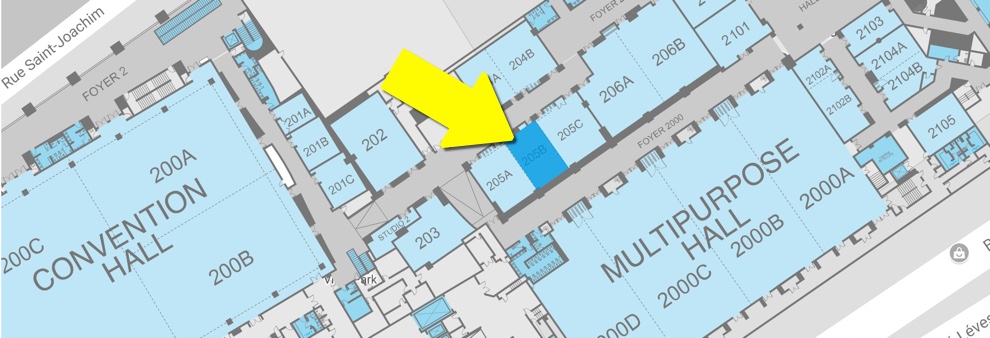
Where: MICCAI 2017, Quebec Convention Center, Room 205B

DICOM4MICCAI is a new tutorial that we presented at the MICCAI 2017 conference. All of the materials (slides, software, datasets, instructions) are accessible following the links below. Please give it a try! If you have any suggestions or feedback, you can use the form at the bottom of this page to let us know how we did, and what we can do better!

The objective of this tutorial is to introduce the MICCAI community to the new kinds of DICOM objects and capabilities that can be used for storage and communication of the data typically produced in the process of quantitative image analysis.

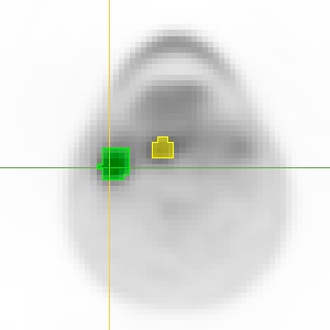
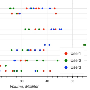
Over the course of presentations and hands-on sessions, we explain how DICOM can, and perhaps should, be used for storing your processing results such as segmentations, parametric maps and volumetric measurements.

After completing this tutorial, attendees are expected to develop an understanding of the relevant new capabilities of the DICOM standard, as well as some of the tools that they can use to experiment with adoption of the standard in their everyday research.

Schedule

  • 1:30pm

    Introduction, goals, scope

  • 1:40pm

    DICOM: Overview and historical perspective - David Clunie

    This talk will introduce the main concepts of the DICOM standard, the role it plays in the modern healthcare enterprise, its overall purpose and capabilities.

    Slides - PDF

  • 2:10pm

    DICOM for Quantitative Imaging - Andrey Fedorov

    This talk will discuss the relevance of the DICOM standard in quantitative imaging research, motivate its use and discuss specific capabilities as applied to the storage of common image analysis outputs, such as image segmentation results, parametric maps and region-based quantitative measurements.

    Slides - PDF

  • 2:35pm

    Standardizing on DICOM for oncology use cases - Marco Nolden

    This presentation will discuss how the concepts and capabilities of DICOM standard can be utilized to support quantitative imaging in cancer research applications.

    Slides - PDF

  • 3:00pm

    Hands-on tutorial prerequisites setup

    Attendees will be guided over the steps of installing the free open source software tools that will be used in the tutorial, and which can help the attendees work with the DICOM data in their everyday work after the conference. The tools utilized in the tutorial will include 3D Slicer, MITK, DCMTK, Atom, and dcmqi. We will also distribute the sample dataset that will be used in the tutorial.

    Follow this link for the instructions on how to install the prerequisites for your platform in advance of the tutorial!

  • 3:30pm

    Coffee!

    The specific roast of coffee that will be served is to be finalized by the MICCAI 2017 organizers, but we promise it will be free (considering you paid the registration fee) ;-)

  • 4:00pm

    Hands on session: Using DICOM to store your analysis results

    Starting with an example of image segmentation task, we will demonstrate how 3D Slicer can be used to segment a DICOM image, calculate quantitative measurements over the segmented regions, and store the result using DICOM Segmentation object and DICOM Structured Report. We will then explore the content of the resulting objects, and demonstrate how they can be interpreted using MITK. If there is time left, we will continue with the demonstrations of how to convert legacy analysis results generated using non-DICOM-aware tools into the standard format, and how to convert quantitative image analysis results stored in DICOM into popular research formats.

    Tutorial materials

  • 4:45pm

    Hands on session: Exploring data stored in DICOM

    Starting with a relatively large publicly available DICOM dataset that contains PET and CT image data together with multiple segmentations of various structures and quantitative measures extracted from those, we will demonstrate how to convert DICOM data into a tabular form suitable for analysis, and will use Jupyter notebook and related python data analysis tools to explore various types of data present in DICOM.

    Tutorial materials

Speakers

We look forward to meeting you!

David Clunie

Radiologist, medical informaticist, DICOM open source software author and editor of the DICOM standard. Formerly the co-chair of the IHE Radiology Technical Committee and industry co-chairman of the DICOM Standards Committee.

Andrey Fedorov

Assistant Professor in Radiology at the Brigham and Women's Hospital Department of Radiology, and Harvard Medical School. Together with Ron Kikinis, Andrey is a co-PI of the Quantitative Image Informatics for Cancer Research project.

Marco Nolden

Medical Imaging Interaction Toolkit (MITK) team lead at the German Cancer Research Center. His work focuses on research enabling technologies, interoperability, open source and application-oriented research in radiology, surgery and radiation oncology.

Feedback

Your feedback can help us improve the tutorial materials, and do a better job next time we teach it in person!